The Body Held in Tension
Fascia isn't packing material — it's a tensioned net suspending the skeleton, and its cables are mostly water. A working tensegrity model, from Levin and Ingber's biotensegrity to the hydration that actually governs it.
Drag any point — the whole net resists, then recoils.
Levin's spine mechanics, Ingber's cytoskeleton, the hydration that actually drives it — and where the model holds up, or doesn't.
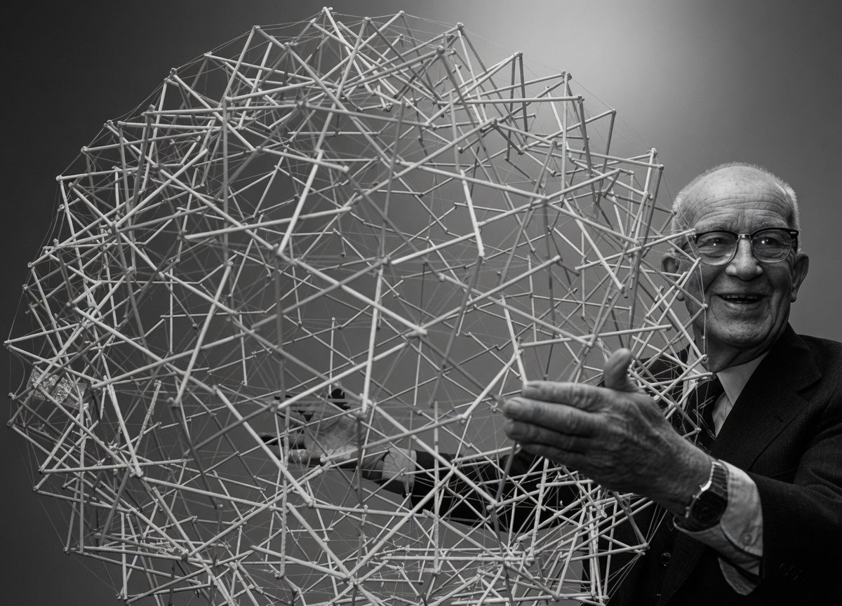
For most of modern medicine, the skeleton has been treated as scaffolding. Bones stack, joints hinge, and everything else — muscle, tendon, and the thin white webbing called fascia — is described as padding, wrapping, plumbing. Structural, but secondary. A frame with furniture on it.
Tensegrity says the frame is wrong.
The word is a 1955 coinage of Buckminster Fuller’s, welded from “tension” and “integrity.” He didn’t build the first one. That was Kenneth Snelson, a student in Fuller’s 1948 summer class at Black Mountain College, who went home to Oregon that winter and came back with a sculpture unlike anything his teacher had shown him: rigid struts suspended in mid-air, never touching, held in a fixed and startlingly strong configuration by wires alone. Fuller was captivated, named the principle, patented broad versions of it, and — depending on whose account you read — spent the following decades not crediting the twenty-two-year-old who actually built the thing. Snelson kept sculpting; his Needle Tower has stood at the Hirshhorn Museum since 1968, sixty feet of aluminum tube that looks like it’s defying gravity because, structurally, it sort of is.
The figure at the top of this page is a body built on that principle. Fourteen struts stand in for bones — the spine’s segments, the clavicle, the pelvis, the limbs — and not one of them stacks on the one below. They don’t touch. What holds the shoulders and hips up, what keeps the whole thing standing, is the surrounding net of tension lines: the model’s stand-in for fascia. It’s pinned only at the feet. Pull the head or a hand and watch what happens — nothing you touch stays local. The tension redistributes across every line at once, the far side of the body leaning to answer a tug on the near side, and when you let go it re-tensions back to standing. There’s no single load-bearing column to buckle, because there is no column. That’s the whole thesis in one object: islands of compression floating in a single continuous net of tension.
The surgeon who saw a spine in it
In 1970, an orthopedic surgeon named Stephen Levin stood on the National Mall in front of one of Snelson’s structures and had the kind of idea that either sounds obvious in hindsight or completely deranged, depending on your training. He’d spent years frustrated with the standard model of the spine — vertebrae as a stack of blocks, discs as cushions between them, load traveling straight down like a pillar under a roof. That model has a problem: real spines bend, twist, and absorb impact in ways a stack of blocks simply doesn’t. Loaded straight down, a stack of blocks buckles or crushes. Spines don’t, not under normal use.
Levin’s alternative, which he later named biotensegrity: the vertebrae are the struts. They never actually bear weight against each other directly — cartilage and fluid keep true bone-on-bone contact vanishingly rare even at the joint surfaces. What holds the column up and lets it move is the continuous wrap of ligament, tendon, and fascia around and through it, in permanent, distributed tension. The spine isn’t a compression stack with tension add-ons. It’s a tensegrity mast, and the “add-ons” are the actual structure.
The cell before the skeleton
A parallel argument was building at the same time from an entirely different direction — inside single cells. Donald Ingber, then a graduate student, had been folding paper-and-elastic tensegrity models as a hobby when he noticed the ones he built looked exactly like micrographs of a cell’s cytoskeleton: microtubules as struts, holding their shape only because actin filaments and the cell membrane held them in tension, none of it touching in a rigid frame. Ingber went on to show that cells mechanically stiffen, soften, and change shape according to tensegrity rules — that squeezing a cell from one side changes tension clear across it, instantly, the same way pulling one node of the model above moves every cable in the net.
That’s the claim biotensegrity makes at every scale: the same tension-and-floating-compression logic, repeating from the cytoskeleton up through the fascial web that laces around every muscle, organ, and bone, up to the whole standing body. Not a skeleton with tissue draped over it. A continuous fabric of tension with islands of compression — bones — floating inside it, most of them never truly bearing load against their neighbors at all.
The wires are mostly water
Snelson’s cables are steel. The body’s are not — they’re a hydrogel, and that difference turns out to matter more than the geometry does.
Fascia’s tension-bearing matrix — the “ground substance” it’s suspended in — is built from long chains of glycosaminoglycans, chiefly hyaluronan, which bind water at many times their own weight and hold it there. That water isn’t incidental packing. It’s what lets adjacent fascial sheets glide across each other rather than stick. The French hand surgeon Jean-Claude Guimberteau spent years filming inside living, unanesthetized tissue during hand surgeries and found not the tidy layered sheaths in anatomy textbooks but a continuous, disordered froth of tiny fluid-filled chambers — he called them the “multimicrovacuolar system” — sliding past one another wherever the tissue moved. Dehydrate that froth and the sliding stops; the layers that should glide start to shear and drag against each other instead, which is a large part of what “tight fascia” actually is at the tissue level.
The fluid doesn’t sit still, either. It’s continuous with the interstitium — the fluid-filled space between cells, once treated as a passive gap and reclassified in a widely covered 2018 Scientific Reports paper as a structured, drainable network of collagen-bundle-walled channels running beneath skin, around organs, and through fascia itself, feeding directly into the lymphatic system. Water moves into fascia from the capillary beds it wraps, saturates the ground substance, drains onward through that interstitial network toward lymph, and gets replenished by movement and pressure changes as much as by anything circulatory. Fascia researchers like Robert Schleip have argued this hydration cycle, not collagen density, is the more clinically useful lever: fascia that moves regularly stays hydrated and slides; fascia that doesn’t stiffens and adheres, independent of how much collagen is actually there.
Which is the point the tensegrity model can’t show you, because its cables can’t get thirsty. A Snelson sculpture has one tension state — the wires are the length they are. A body’s tension net has a variable the sculpture doesn’t: how hydrated its cables are on a given day, in a given tissue, changes how that tension actually behaves — stiffer and stickier when dry, more freely sliding and evenly load-sharing when not. The strut-and-cable geometry explains how force distributes through the net. The water explains why the same net can feel like a completely different structure from one week to the next.
The interstitium-as-organ framing is itself disputed — critics note the spaces Benias and colleagues described were largely already known to anatomists, and “new organ” may oversell a reclassification of existing structure. What’s not seriously contested is the underlying finding: fascia is a fluid-perfused, actively draining tissue, not a static membrane, and its mechanical behavior tracks its hydration state closely enough that clinicians treat “get the water moving” as a real intervention, not a wellness slogan.
What actually follows from this
The honest version of this story includes real disagreement. Biotensegrity is a contested framework, not settled anatomy — critics point out that real joints, unlike an idealized tensegrity module, do transmit substantial compressive load through direct contact (that’s what cartilage is for), and that “everything is tensegrity” can become an unfalsifiable metaphor if you’re not careful about which claims are structural fact and which are analogy. Fascia researchers who take the model seriously tend to hold a narrower, more defensible version of it: that fascia is not inert wrapping, that it forms one mechanically continuous network rather than isolated sheaths, and that force applied anywhere in the body measurably transmits through that network rather than staying local — a claim with real electromyography and cadaver-dissection evidence behind it, distinct from the stronger claim that bones never bear compressive load at all.
What survives the disagreement is the shift in what you’re looking at when you look at a body. Not a frame with soft parts attached, but a tensioned net with hard parts floating inside it — same logic as the model above, same logic as Snelson’s aluminum tower, applied at a scale small enough to walk around in.